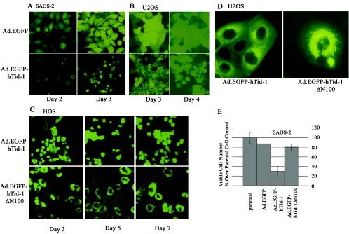
FIG. 8.
Adenovirus-mediated gene transfer of hTid-1 leads to cell death in a J domain-dependent manner. Recombinant adenoviruses expressing EGFP (control) and EGFP-tagged hTid-1 were used to transduce SAOS-2 (A) and U2OS (B) cells. At the indicated times, fluorescent images were recorded. (C) HOS cells were transduced with recombinant adenoviruses expressing EGFP-tagged full-length hTid-1 and the hTid-1 ΔN100 mutant. The fluorescent images were taken at different times, as indicated in the figure. The results represent at least three independent experiments. (D) Digitally enlarged fluorescent images show the subcellular distribution of EGFP-hTid-1 and EGFP-hTid-1ΔN100 in transduced U2OS cells. (E) In vitro cytotoxicity assay to assess the viability of SAOS-2 cells transduced with the recombinant adenoviruses expressing EGFP (control), EGFP-hTid-1, and EGFP-hTid-1ΔN100. At 3 days after transduction, viable cells were counted with a microscope, and dead cells were excluded by trypan blue staining.

