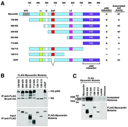
FIG. 4.
Mapping of the region of myocardin that interacts with p300. (A) Schematic diagram of myocardin and the mutant forms used to map the p300-binding domain. NT, not tested. (B) Coimmunoprecipitation assays. COS cells were transiently transfected with expression vectors encoding FLAG-tagged myocardin deletion mutant proteins and hemagglutinin (HA)-tagged p300. Myocardin proteins were immunoprecipitated (IP) from cell lysates with a monoclonal anti-FLAG antibody, and coimmunoprecipitating HA-tagged p300 was detected by immunoblotting (IB) with a polyclonal anti-HA antibody (top part). The expression of myocardin proteins was revealed by an anti-FLAG antibody (bottom part). NS, nonspecific background. (C) COS cells were transiently transfected with expression vectors encoding the indicated myocardin deletion mutant proteins, and immunoprecipitates were assayed for HAT activity in the presence of purified histones and 3H-labeled acetyl coenzyme A, followed by SDS-PAGE, as described in Materials and Methods.

