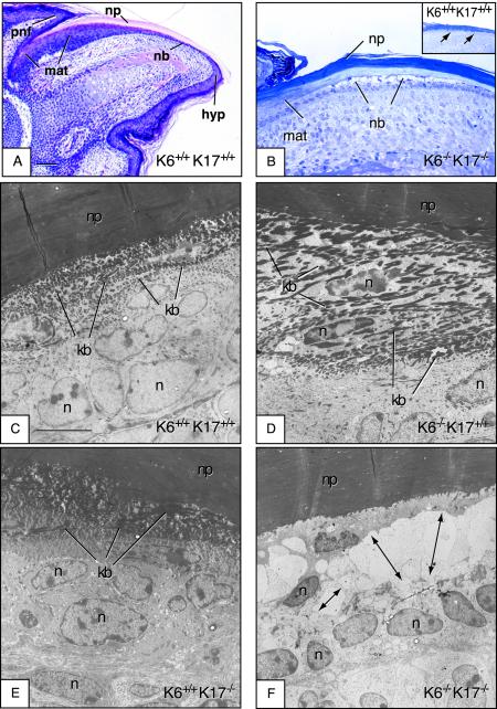
FIG.2.
Early lysis of the nail bed epithelium in K6α/K6β−/− K17−/− null newborn mice but not in other genotypes. Paws or digits from P0 mice (newborns) were surgically removed and processed for routine histology. Micrographs shown are derived from an H&E-stained section of paraffin-embedded tissue (A), toluidine-blue stained semithin sections (∼0.5 μm thick) prepared from epoxy-embedded tissues (B), and uranyl acetate- and lead citrate-counterstained stained ultrathin sections (∼50 to 70 nm thick) prepared from epoxy-embedded tissue (C to F). In all cases, the sectioning plane is along the anterior-posterior axis and the mouse genotype is indicated in the lower right corner. (A) Section of wild-type nail tissue showed for orientation. The proximal nail fold (pnf), matrix (mat), nail bed (nb), nail plate (np), and hyponychium (hyp) compartments are depicted. Bar, 300 μm. (B) Semithin section of epoxy-embedded K6α/K6β−/− K17−/− nail unit, depicting the nail bed area. There is striking lysis of the nail bed (nb) epithelium, while the matrix (mat) and nail plate (np) appear not affected. The inset shows an identical preparation from a wild-type mouse, which does not show any nail bed (nb) alteration. (C to F) Electron microscopy data taken from a smaller subset of the same general area (nail bed). In contrast to K6α/K6β+/+ K17+/+ (C), K6α/K6β−/− K17+/+ (D), and K6α/K6β+/+ K17+/+ (E) mice, the nail bed of K6α/K6β−/− K17−/− mice (F) shows obvious ballooning and lysis in the area immediate above the basal layer (see arrows). Likewise, the two to three layers of cells with dense keratin bundles (kb in panels C to E) are missing from the K6α/K6β−/− K17−/− sample (F). In contrast, the nail plate (np) and matrix appear normal in these mice. n, nucleus. Bar, 5 μm (C to F).

