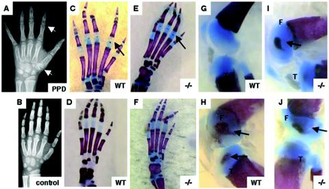
FIG. 4.
Secondary centers of ossification in wild-type (WT) and Wisp3−/− mice. (A and B) Hand radiographs of an 8-year-old patient with PPD showing enlarged epiphyses within phalanges and carpal bones (white arrows) compared to an age- and gender-matched control. (C to J) Alcian blue-stained (cartilage) and alizarin red-stained (bone) 7-day-old wild-type and Wisp3−/− mouse hind paws (C to F) and knee joints (G to J) do not reveal genotype-specific effects on secondary centers of ossification. Black arrows indicate epiphyseal centers. F, femoral condyle; T, tibial plateau. Magnification, ×8 (G to J) and ×10 (C to F).

