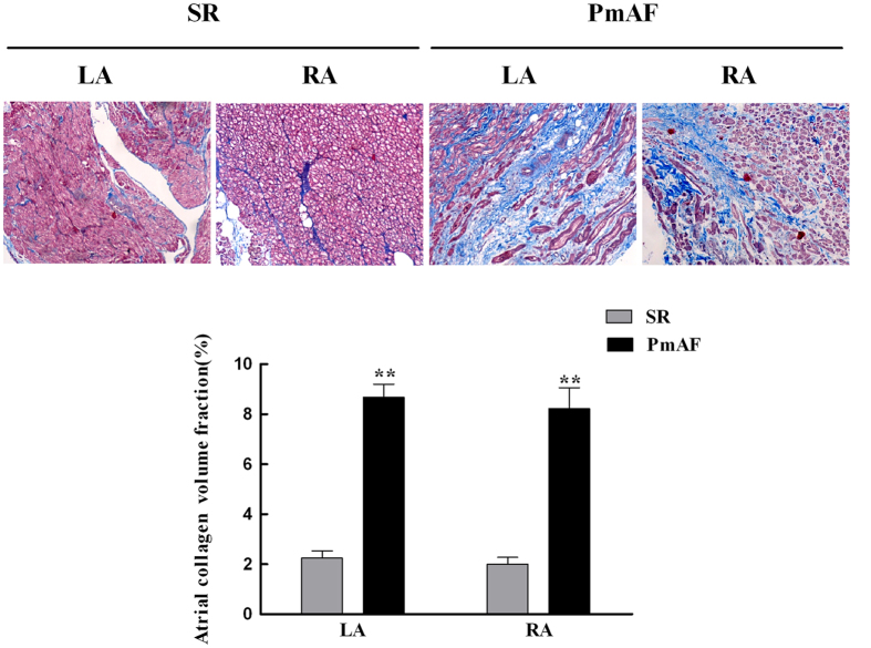
Figure 2. Histopathological changes in atrium of patients.
Atrial collagen determined by Masson staining. Atrial collagen in the right and left atrium of patients with PmAF were significantly higher than in patients with SR. Quantification of atrial collagen is shown. SR group n = 23, PmAF group n = 19, **P < 0.01 compared with the SR group. SR = Sinus rhythm; PmAF = Permanent AF; LA = left atrium, RA = right atrium. Magnification: ×100.

