FIG. 2.
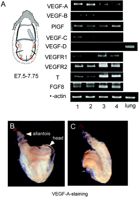
Spatially distinct expression pattern of genes encoding the VEGF ligand and receptors in embryos from E7.5 to E7.75. (A) Embryos were divided into four parts (left), and the mRNA level in each part was semiquantitatively estimated by RT-PCR (right). RNA derived from the lung was a positive control for VEGF-D. (B and C) Immunohistochemistry showing the distribution of VEGF-A protein in front (B) and side (C) views of a whole embryo. The VEGF-A signal was stronger in the anterior than the posterior portion of the trunk.

