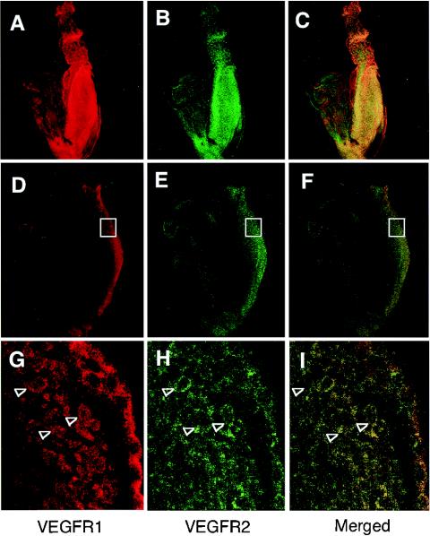
FIG. 3.
Posterior cells dominantly express both VEGFR1 and VEGFR2. Sagittal sections of embryos at E7.75 were stained with anti-mouse VEGFR1 (A, D, and J) or anti-mouse VEGFR2 (B, E, and H), and the images were merged (C, F, and I). The embryo was scanned by confocal microscopy, and a three-dimensional image was reconstructed to represent the whole embryo (A to C). The colocalization of VEGFR1 and VEGFR2 in posterior cells (open boxes in panels D to F) was apparent at a higher magnification (arrowheads in panels G to I).

