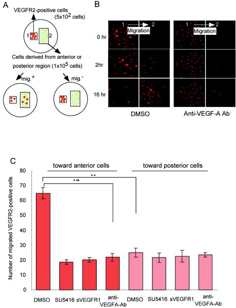
FIG. 5.
VEGFR-positive cells move toward the anterior portion of embryos with concentrated VEGF in vitro. (A) Scheme of the assay system enabling the migration of VEGFR-positive cells toward embryonic cells derived from the anterior or posterior portion of several wild-type embryos at E7.5. (B) Rhodamine-labeled VEGFR-positive embryonic cells migrated toward cells from the anterior region after 0, 2, and 6 h (left panels). A neutralizing anti-mouse VEGF-A antibody strongly suppressed the migration of the cells derived from the anterior portion (right panels). The direction of migration was from box 1 to box 2. The images were made by dark-field microscopy. (C) Numbers of migrated VEGFR2-positive cells in medium containing DMSO as a negative control, 10 μM SU5416, 1 μg of sVEGFR1/ml, or 1 μg of neutralizing anti-mouse VEGF-A antibody/ml. *, P < 0.05 by a t test.

