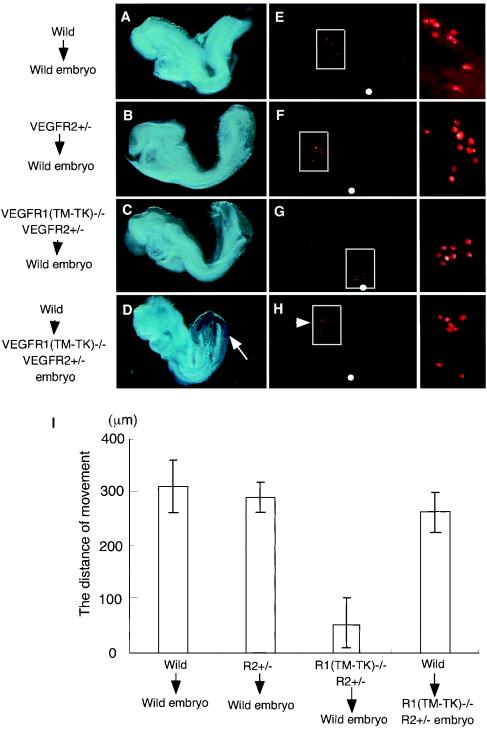
FIG. 6.
VEGFR-positive cells migrate in the anterior direction in a manner dependent on VEGFRs in vivo. (A to H) Colonization of rhodamine-labeled VEGFR-positive cells derived from wild-type (A and E), VEGFR2+/− (B and F), and VEGFR1(TM-TK)−/− VEGFR2+/− (C and G) mice in wild-type mice. (D and H) Transplanted cells from wild-type mice migrated to the cranial region (arrowhead) compared to host VEGFR2-positive cells (arrow) in VEGFR1(TM-TK)−/− VEGFR2+/− embryos. The left panels show a bright field view, and the middle panels show a dark field view. The boxed part of the dark field view is magnified in the right panels. (I) Distances migrated from the injection site (white circle in panels E to H). More than 90% of the VEGFR-positive cells derived from VEGFR1(TM-TK)−/− VEGFR2+/− mice remained around the injection site.

