FIG. 6.
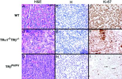
Ki67 Immunohistochemistry in pituitaries of wild-type, TRβPV/PV, and TRα1−/− TRβ−/− mice. Immunoperoxidase labeling for Ki67 was performed on pituitaries from wild-type (A to C), TRα1−/− TRβ−/− (D to F), and TRβPV/PV (G to I) mice. Panels A, D, and G are stained with hematoxylin and eosin and panels B, E, and H are stained with hematoxylin. Panels A, B, D, and E showed no evidence of adenomas; whereas panels G and H show adenoma cells with enlarged nuclei. Panels C and F show a low level of cell proliferation in wild-type and TRα1−/− TRβ−/− mice, respectively. The anti-Ki67 antibody used showed a background nonspecific cross-reaction with a granule antigen in nonthyrotrophs (C and F), but the level of nuclear labeling was very low (arrowheads). In contrast, nuclear labeling for Ki67 was high in TSH-oma areas (arrows) (6.1% of all nuclei). Bar, 40 μm.

