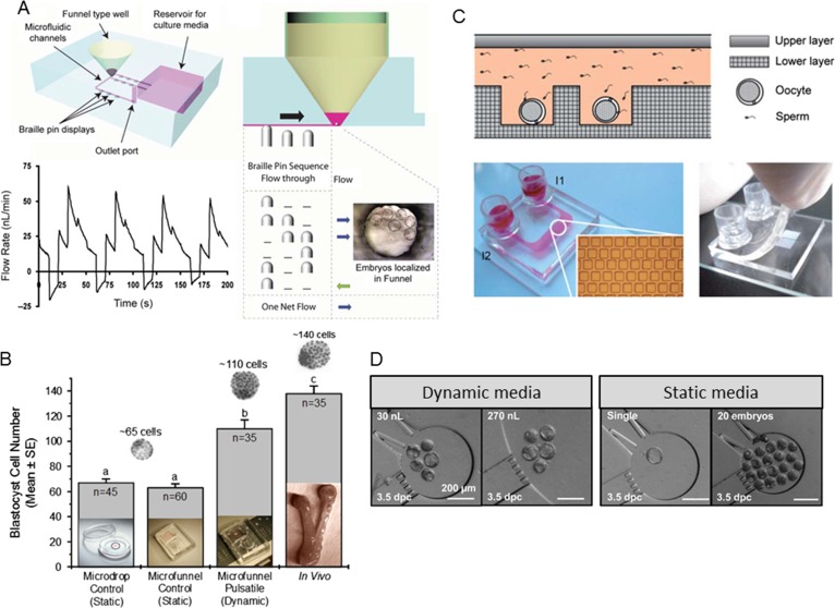
Figure 2.
(A) Schematic drawing of a microfunnel cartridge used for culturing mouse embryos. The cartridge is placed on an array of piezoelectric pins with actuation provided by Braille pins. Embryos are placed into media, overlaid with oil, within microfunnels under the flow-through condition created by the pin actuation sequence. The flow pattern generated over four cycles of the five-step Braille pin actuation sequence at 0.1 Hz. (B) Dynamic fluid condition (microfunnel-pulsatile) shows a greater number of cells per blastocyst compared with static culture (in either media microdrops under oil or media in microfunnels under oil with no flow) within the same amount of culture time, with results closer to in vivo conditions (when flushed out of the uterus at a time corresponding to culture length; a,b,c = P < 0.01). Adapted from Heo et al. (2010). (C) Illustration of the cross-section of the double-layer microfluidic device used for culturing mouse embryos. A view of the microfluidic device fixed on a 76.2 mm × 25.4 mm microscopic slide, as well as the microwell array micrograph. The microchannel and the two reservoirs were filled with red dye. Scale bar is 500 mm. Finally, a photograph of the microfluidic device with the upper layer lifted. Adapted from Han et al. (2010). (D) Micrographs of mouse embryos cultured in nL chambers under various conditions. Adapted from Esteves et al. (2013).

