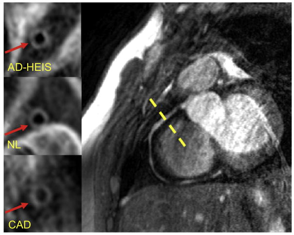
Fig. 1. Representative MR images of coronary vessel wall imaging.
Example of right coronary artery 3T MRI image. Vessel wall image for one of the subjects in each of the three groups is shown in the left panels. AD-HIES, autosomal dominant hyper-IgE syndrome; NL, healthy subject; CAD, coronary artery disease.

