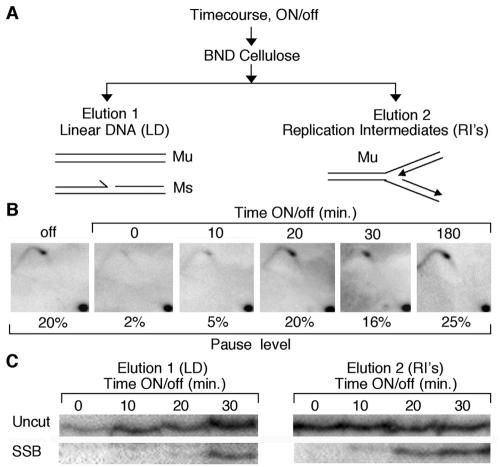
FIG. 4.
The SSB and MPS1 occur simultaneously. (A) Schematic showing how linear DNA and replication intermediates are enriched from purified time course DNA. (B) A thiamine time course was performed from isolated virgin Mu cells, and DNA harvested from cells was analyzed by 2D gel and PAGE (C, right panel), to monitor the kinetics of the MPS1 pause and imprint or SSB formation. Upon addition of thiamine (nmt1 repression), the pause structure and break are formed at the same time, within 10 min. The percentage of pausing is indicated (C). Denaturing PAGE gel analysis of the SSB shows that it is part of a replicating structure. Linear DNA (LD) and replication intermediates (RI’s) from the same time course DNA used in the results shown in panel B were collected and analyzed by denaturing PAGE. The SSB is first seen in the replication intermediate-containing fraction (elution 2).

