
Figure 2.
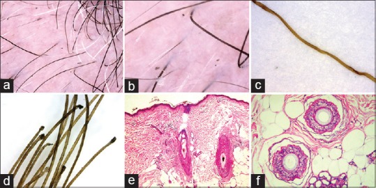
Trichoscopic and histopathologic findings in patients treated with erlotinib. Trichoscopy shows (a) black dots, twisting of hair shafts, and broken hairs at different lengths (×20); (b) features resembling black rectangular granular structures (×20); (c) high magnification of a flattened hair shaft reveals multiple twisting (×250); (d) the pull test results in numerous anagen hairs devoid of sheaths (×250); (e) scalp biopsy from the alopecic area shows two hair follicles corresponding to the broken hairs on trichoscopy as their hair shafts are replaced by pigmented casts in the hair canal and are surrounded by abnormal, disintegrated inner root sheath, and irregularly thinned outer root sheath. Note the absence of sebaceous glands (H and E, vertical sections, ×4); (f) two anagen follicles at the level of subcutaneous fat reveal corrugated and thicker pink vitreous layer (H and E, horizontal sections, ×10)