Figure 2.

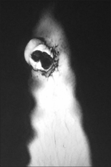
Magnetic resonance imaging of the skin lesion. Well-demarcated mass with centrally solid and peripherally cystic structures

Magnetic resonance imaging of the skin lesion. Well-demarcated mass with centrally solid and peripherally cystic structures