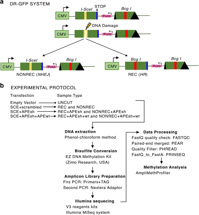
Figure 1. DRGFP system and the experimental protocol.
(a) Schematic diagram of the DRGFP system. I-SceI (yellow line) indicates the site cleaved by the meganuclease I-SceI. The I-SceI site is converted by HR into a new site for the enzyme BcgI (Bcg I red line). The first and the second cassettes are shown. Cassette II is not transcribed. (b) Schematic representation of the experimental protocol. HeLa DRGFP cells were transfected with (1) SCE+scrambledshRNA; (2) SCE+APEsh; (3) SCE+APEsh+APEwt (on the left). Recombinant (REC) and non-recombinant or NHEJ (NONREC) molecules were purified following each transfection. UNCUT represents control plasmid-transfected HeLa DRGFP cells. The arrows indicate the steps and the procedures undertaken in analyzing the DNA methylation data.

