FIG. 10.
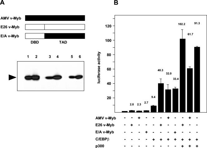
Cooperation of C/EBPβ and p300 with different v-Myb variants. (A) A schematic illustration of different forms of v-Myb is shown at the top. The bottom shows a Western blot of QT6 cells transfected with 3 (lanes 1, 3, and 5) or 5 (lanes 2, 4, and 6) μg of the indicated expression vector stained with Myb-specific antibodies. DBD, DNA-binding domain; TAD, transcription activation domain. (B) QT6 cells were transfected with 2 μg of the reporter gene containing mim-1 enhancer and mim-1 promoter (mim3mim-Luc), pCMVβ (0.2 μg), and expression vectors for C/EBPβ (0.2 μg), p300 (5 μg), E26 v-Myb (1 μg), AMV v-Myb (1 μg), or E26 v-Myb and AMV v-Myb (E/A v-Myb) (1 μg), as indicated at the bottom. Controls contained equivalent amounts of empty expression vector. Luciferase and β-galactosidase activities were determined 24 h after transfection. Bars show the average luciferase activity normalized with respect to the β-galactosidase activity and expressed in arbitrary units. The T bars show standard deviations. The luciferase activity in the absence of exogenous factors was designated 1.

