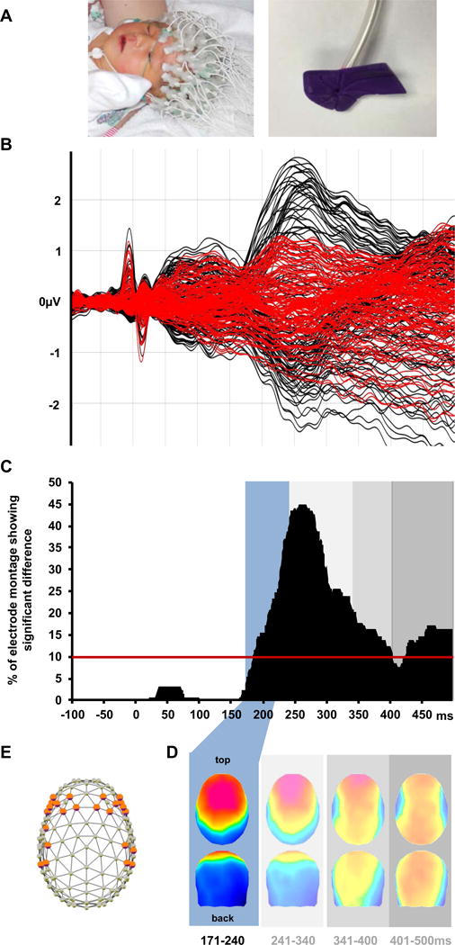
Figure 1.

Normative analysis of ERPs from full-term infants. A. Photos of a full-term infant undergoing EEG recording (left) and the tubing and nozzle for delivering calibrated light touch to the hand (right). B. Superimposed ERPs to touch and sham stimuli (black and red traces, respectively). C. Significant ERP differences began at 184ms post-stimulus onset (percentage of significant electrodes across time shown). D. Hierarchical topographic clustering identified a series of touch-related ERP components (shaded boxes); the earliest, 171–240ms, was the focus of the present analyses. e. 24 bilateral electrodes were at the maxima/minima of the blue ERP topography, and measures from these were used in subsequent analyses. For term patient characteristics see Table S1.
