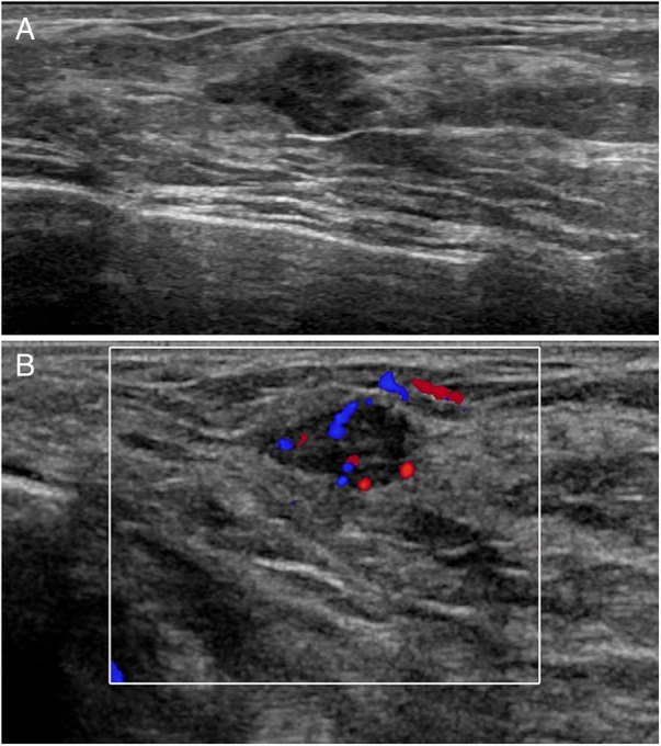
Figure 2.

Ultrasound images: (A) there is an irregularly shaped, hypoechoic mass with indistinct margins (B) colour Doppler shows prominent internal vascularity (Doppler settings: 77% saturation, 487 Hz, wall filter 26 Hz, pulse repetition frequency (PRF)±2.5 cm/s).
