
FIG. 2.
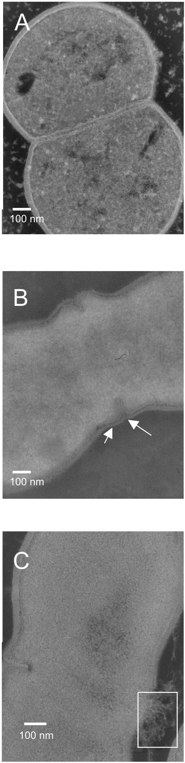
Effect of d-Ala depletion on the cell integrity. (A) Electron microscopy image of L. lactis NZ3900 in stationary phase. (B) Electron microscopy image of L. lactis PH3960 (Δalr) after 5 h of d-Ala depletion. Arrows show the thinner cell wall around the septum. (C) Electron microscopy image of L. lactis PH3960 (Δalr) after 2 h of d-Ala depletion. The white rectangle shows where release of cytoplasmic material occurs.