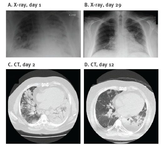
Figure 1.
Chest X-ray and computed tomography in a patient with severe swine influenza A(H1N1), Italy, October 2016
CT: computed tomography.
The X-ray taken on admission (A) showed bilateral opacities. A follow-up chest X-ray performed on day 29 showed almost complete regression of the opacities (B). Chest CT on day 2 after admission (C) showed extensive bilateral consolidation with alveolar parenchymal consolidations and ground-glass opacity (right end). CT on day 12 after admission showed partial regression of consolidation in the right median-lower lobe with residual little areas of ground-glass opacity and improved ventilation of the lower left lobe (D).

