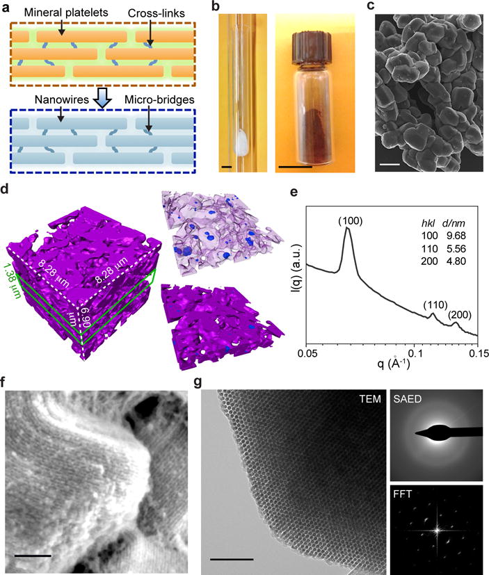
Figure 1. Amorphous Si can have multi-scale structural heterogeneity and ordered mesoscale features.

a, Si-based cellular modulation materials can be designed to share similar mesostructures with natural biomaterials, e.g., bones, with ordered unidirectional fibril networks that are maintained by molecular cross-links. b, A double quartz-tubing system was used for nano-casting synthesis, with a mesoporous SiO2 template placed near the bottom of the inner tube (left). After HF etching, the brownish product (right) can be yielded. Scale bars, 1 cm. c, SEM image of as-synthesized Si particles shows a morphology similar to that of SBA-15 template. Scale bar, 2 μm. d, TXM 3D dataset of a representative region of mesostructured silicon (left). A thin slice of the dataset (green lines) highlights the presence of both intra- and inter-granular voids (right). Representing silicon as a semi-transparent matrix allows clearer visualization of the voids (upper right). Magenta, silicon; blue, intra-granular voids; open regions in the whole volume or thin slice, inter-granular voids. e, SAXS profile shows mesoscale periodicity with a 2D hexagonal symmetry. f, SEM image reveals periodic arrangement of Si nanowire assembly. Scale bar, 100 nm. g, TEM image (left) and its FFT diffractogram (lower right) indicate the hexagonal packing of Si nanowires (left panel). SAED pattern shows an amorphous atomic structure (upper right). Scale bar, 100 nm.
