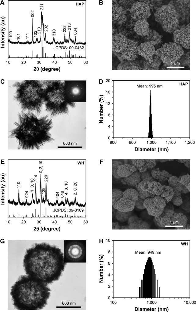
Figure 1.
XRD patterns (A and E), SEM micrographs (B and F), TEM micrographs (C and G) and DLS size distributions (D and H) of HAP (A–D) and WH (E–H) porous hollow microspheres that were prepared by using creatine phosphate disodium salt as an organic phosphorus source through the microwave-assisted hydrothermal method at 120°C for 10 min. Insets of (C) and (G) are selected-area electron diffraction patterns.
Abbreviations: XRD, X-ray diffraction; SEM, scanning electron microscope; TEM, transmission electron microscopy; DLS, dynamic light scattering; WH, whitlockite; HAP, hydroxyapatite.

