
Figure 2.
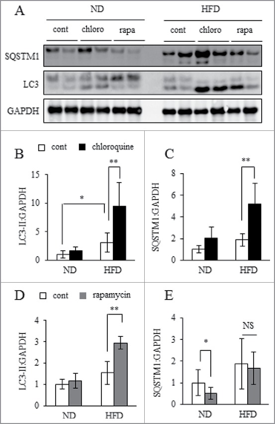
Autophagosome formation was accelerating but lysosomal clearance was impaired. WAT explants from ND or 30HFD mice were incubated in the presence or absence of 10 mM chloroquine (chloro) or 500 nM rapamycin (rapa) for 24 h, and then samples were collected and assayed. Total protein extracted from WAT was analyzed by western blot using anti-SQSTM1, LC3 and GAPDH antibodies (A) with quantitative data shown (B to E). Representative images and the quantitative data (n = 4) were shown. Intensity of GAPDH was used as a loading control. Values indicate mean ± SD (n = 6). Differences between values were analyzed by the Student t test. Statistical significance shown as *P < 0.05, **P < 0.01.