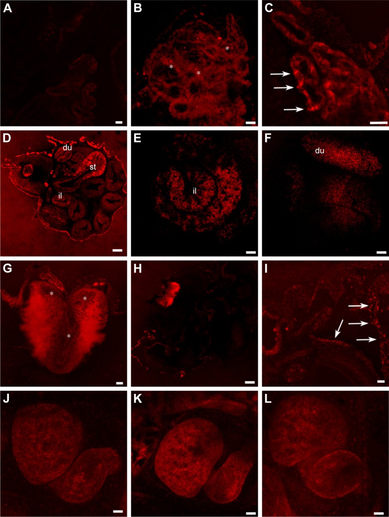
Figure 8.
gH625-QDs confocal localization in stage 46 Xenopus laevis embryos.
Notes: (A) Control section. (B, C) Optical sections of primordium of lung treated with (B) naked QDs and (C) gH625-QDs. QDs localize in the form of widespread dots (B, asterisks) in contrast gH625-QDs, which are disposed in small fluorescent areas (C, arrows). In the intestine (D–F), gH625-QDs are visible only in some stretches: st, du and il. Hindbrain shows a slight localization of gH625-QDs (G, asterisks). (H) QDs are barely visible in the gills (I) gH625-QDs have a distribution similar to the gills (arrows). (J–L) QDs and gH626-QDs are not visible in the heart. Bars: (A–C, G, I) =20 µm; (E, F, K, J, L) =50 µm; (H) =100 µm; (D) =200 µm.
Abbreviations: du, duodenum; il, ileum; QDs, quantum dots; st, stomach.

