FIG. 5.
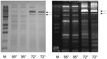
SDS-PAGE analysis of membrane fractions of cold-adapted cells stained for carbohydrates. Results are shown for the SDS-PAGE (15%) analysis of the salt-washed (4.0 M NaCl) membrane fractions of P. furiosus cells grown at the indicated temperature (72 or 95°C). M, marker proteins. The gels were stained for protein (with Coomassie brilliant blue) (left) or for carbohydrate (with Emerald 300 Q glycoprotein stain) (right). The arrows indicate the positions of the bands representing CipA (upper) and CipB (lower).

