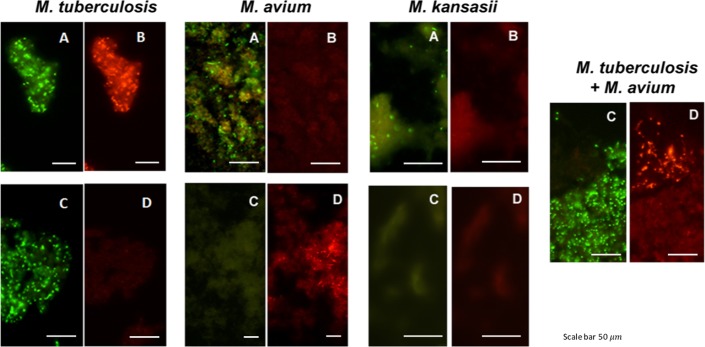
Fig 1. Photographs showing the dual color fluorescence reactivity of individual cultures of Mycobacterium tuberculosis, Mycobacterium avium and Mycobacterium kansasii in the MN Genus-MTBC FISH and MTBC-MAC FISH assays and of a mixed culture of Mycobacterium tuberculosis and Mycobacterium avium in the MTBC-MAC FISH assay.
A, B–Dual fluorescence reactivity in the same microscopic field with the MN Genus- and MTBC-specific probes respectively in the MN Genus-MTBC FISH assay; C, D—Dual fluorescence reactivity with the MTBC- and MAC-specific probes respectively in the MTBC-MAC FISH assay. Mycobacteria used in the FISH tests were: LJ culture of M. tuberculosis (ATCC 25177); LJ culture of M. avium (ATCC 25291); MGIT culture of M. kansasii (ATCC 12478); and an artificially mixed LJ culture of M. tuberculosis (ATCC 25177) and M. avium (ATCC 25291). Photographs were taken at x1000 magnification. Scale bars shown in the photographs represent approximately 50 μm.

