
Figure 5. Residues S226 and A332 in LITE-1 are required for its absorption of UVA but not UVB light in vitro.
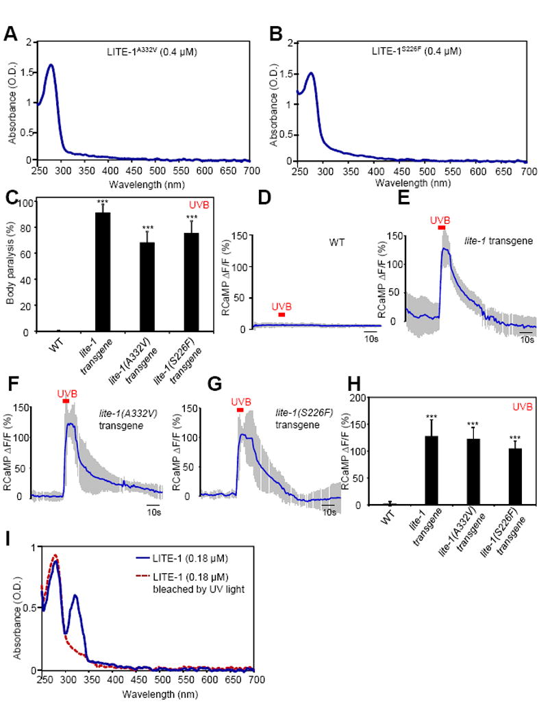
(A–B) S226F and A332V mutations disrupt LITE-1’s absorption of UVA but not UVB light in vitro. The extinction coefficient at 280 nm for LITE-1A332V and LITE-1226F is: 4.0×10⁶ M−1cm−1 and 3.75×10⁶ M−1cm−1, respectively, which are similar to wild-type LITE-1 (Figure 2G).
(C) S226F and A332V mutations do not disrupt the sensitivity of LITE-1 to UVB light in vivo shown by behavioral assays. LITE-1 harboring S226F or A332V was expressed as a transgene in muscle cells under the myo-3 promoter. WT (wild-type) and transgenic worms were exposed to a 20 sec pulse of UVB light (280±10 nm, 0.03 mW/mm2). Animals showing muscle contraction-induced paralysis during light illumination were scored positive. n=20. Error bars: SEM. ***p<0.00001 (ANOVA with Bonferroni test).
(D–H) S226F and A332V mutations do not disrupt the sensitivity of LITE-1 to UVB light in vivo shown by calcium imaging. The experiments were done as described in Figure 2B. A 5 sec pulse of UVB light (280±10 nm, 0.02 mW/mm2) was applied to muscles to elicit calcium transients. Shades along the traces in (D–G) represent SEM. (H) Bar graph. n≥10. ***p<0.00001 (ANOVA with Bonferroni test).
(I) LITE-1 absorption of UVB but not UVA light shows resistance to photobleaching. LITE-1 was pre-exposed to UV light for 5 min (17 μW/mm2, 302 nm) at room temperature prior to spectrophotometric analysis. Pre-exposure to UV light for 30 min still did not notably affect the UVB photoabsorption. The photoabsorption at 280 nm was eventually lost after 1 hour of pre-exposure, probably because LITE-1 was denatured. As a direct comparison, bRho, when tested under the same condition, showed photobleaching of its 568 nm peak in less than 5 min of pre-exposure to ambient light, and such photobleaching became complete at 10 min.
Also see Figure S5.