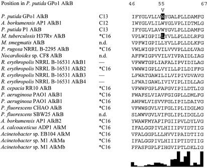
FIG. 3.
Sequence alignment of TM helix 2 and comparison of properties. The first column contains the strain and AH names. M. smegmatis, Mycobacterium smegmatis; P. rugosa, Prauserella rugosa; R. erythropolis, Rhodococcus erythropolis; B. cepacia, Burkholderia cepacia; A. calcoaceticus, Acinetobacter calcoaceticus. The second column contains the upper end of the respective AH substrate ranges. Asterisks indicate that C16 was the longest alkane tested, but it is likely that the substrate range extends to longer alkanes. n.d., not determined; —, no activity could be detected. The third column shows the alignment of TM helix 2, with the position of the first and last residues indicated above (GPo1 AlkB position). TM helix 2 runs from the periplasm to the cytoplasm. Position 55 (GPo1 AlkB position) is indicated by the letter V. The W55 residue in the GPo1 AH and the L69 residue in the M. tuberculosis H37Rv AH are framed in black. The degree of conservation within TM helix 2 is shown below as a bar graph created by Clustal X.

