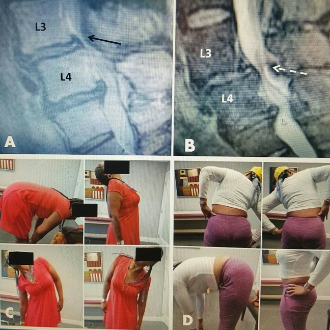
Figure 10. Patient movement after radiofrequency treatment for stenosis.
A: 58-year-old female with marked L3-4 stenosis (black arrow) with anterolisthesis at L3-4 and a small superior L4-5 disc extrusion.
B: Post-treatment MRI follow-up at three months shows reduction in posterior ligamentous stenosis (white dashed arrow).
C: Patient movement at three weeks post RF demonstrating full flexion, extension, and lateral bending.
D: Patient continues with full normal movement six months post RF.
MRI: Magnetic resonance imaging; RF: Radiofrequency.

