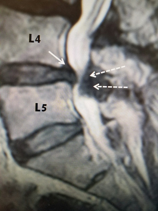
Figure 4. MRI of lumbar spine with L4-5 stenosis.
T2 sagittal MRI of L4-5 stenosis secondary to hypertrophied yellow ligament posteriorly (dashed white arrows) and minimal grade I L4-5 anterolisthesis with disc protrusion posteriorly (solid white arrow). Note that on T2 MRI, bone is a gray color and soft tissue, ligament, and disc annulus are darker.
MRI: Magnetic resonance imaging.

