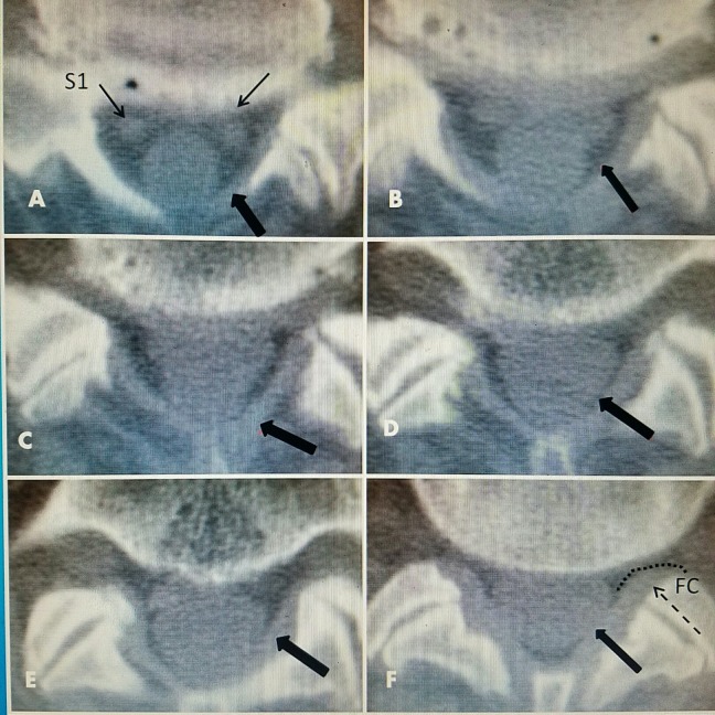
Figure 6. CT axial view of L5-S1 normal ligament flavum.
A-F: Clear delineation of ligamentum flavum (thick black arrows).
A: S1 nerve roots in ventral floor of canal (thin black arrows).
F: Facet capsule (FC) extending laterally at posterior part of neural foramina (dotted line and dashed arrow).
CT: Computed tomography.

