Figure 4.
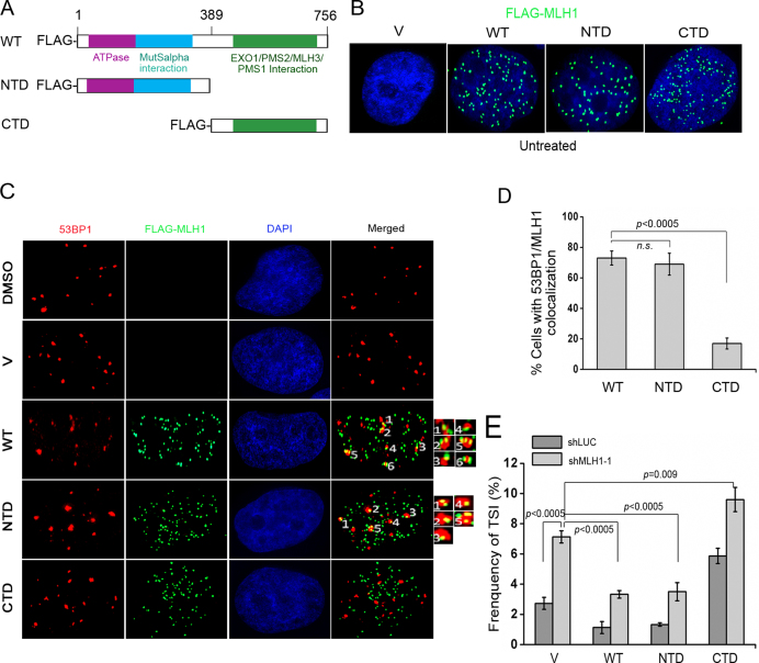
The NTD but not the CTD of MLH1 rescues TSI caused by MLH1 deficiency. (A) Schematic representation of functional domains in MLH1 and its deletion mutants. Purple box: ATPase domain. Blue box: MutSα-interacting domain. Green box: EXO1/PMS2/MLH3-binding domain. (B) Truncated FLAG-MLH1 proteins localize in nucleus in untreated cells. WT and truncated FLAG-MLH1 were stably expressed with retroviral transduction. FLAG antibody (green) was used for IF to detect FLAG-tagged WT MLH1 and mutants. (C) Recruitment of full-length MLH1 (WT) and NTD but not CTD of MLH1 to DNA damage sites induced by etoposide treatment (0.3 μM, 2 h). After etoposide treatment, IF was performed with anti-53BP1 (red) and anti-FLAG (green) in cells expressing WT or mutated FLAG-MLH1. Representative co-localizations of 53BP1 and FLAG-MLH1 were labeled with numbers and then enlarged in insets. (D) Percentage of cells with MLH1/53BP1 co-localization (>5 colocalizations per cell). (E) Frequency of TSI measured in HeLa shLUC and shMLH1 cells with concurrent expression of vector (v), WT-MLH1, NTD, or CTD. Cells were treated with 3 μM etoposide for 2 h and then recovered for 24 h prior to FISH. In each experiment, >1500 chromosomes from each sample were analyzed and each experiment was repeated with three independent replicates.

