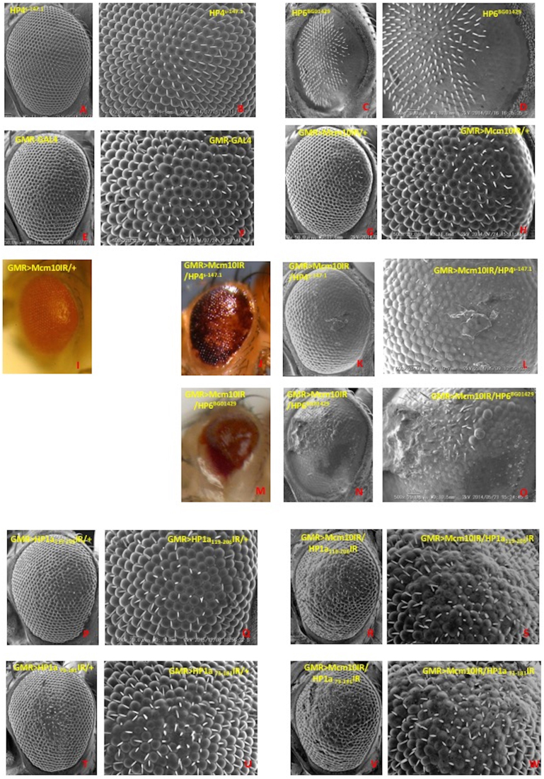
Figure 7.
Scanning electron micrographs and color photo images of adult compound eyes. Posterior is to the right and dorsal is to the top. The flies were reared at 28°C. Panels I, J and M show color photo images and other panels show SEM images. (A and B) w; +; PGSV1-HP4s-147.1, (C and D) w1118; net1 PGT1-HP6BG01429 dpBG01429/ln(2LR)Gla, wgGla-1PPO1Bc, (E and F) GMR-GAL4/+; +; +, (G–I) GMR-GAL4/+; UAS-Mcm10IR/+; +, (J–L) GMR-GAL4/+; UAS-Mcm10IR/+; PGSV1-HP4s-147.1/+, (M–O) w1118; PGT1-HP6BG01429/In(2LR)Gla; +, (P and Q) GMR-GAL4/+; UAS-HP1a119-206IR/+; +, (R and S) GMR-GAL4/+; UAS-HP1a119-206IR/+; +, (T and U) GMR-GAL4/+; +; UAS-HP1a73-181IR/+, (V and W) GMR-GAL4/+; UAS-Mcm10IR/+; UAS-HP1a73-181IR/+. Panels B, D, F, H, L, O, Q, S, U, W (Scale bars indicate 50μm) are higher magnification images of Figure A, C, E, G, K, N, P, R, T, V (scale bars indicate 20 μm), respectively.

