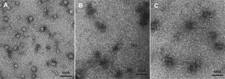
Figure 6.
Compaction of GAL1 RNA by Nab2 ZnF567. (A) Electron micrograph of complexes formed between GAL1 RNA and ZnF567 negatively stained with uranyl acetate following GraFix fixation. Fields contained roughly spherical stain-excluding particles of diameter of the order of 120 Å, consistent with the RNA becoming compacted following binding of the protein. (B) By contrast, rather than excluding uranyl acetate, in the absence of ZnF567 GAL1 RNA bound the stain and so appeared dark as is typically seen with nucleic acids (37). (C) When ZnF567F450A was bound to GAL1 RNA, the particles were less compact than those obtained with the wild-type protein.

