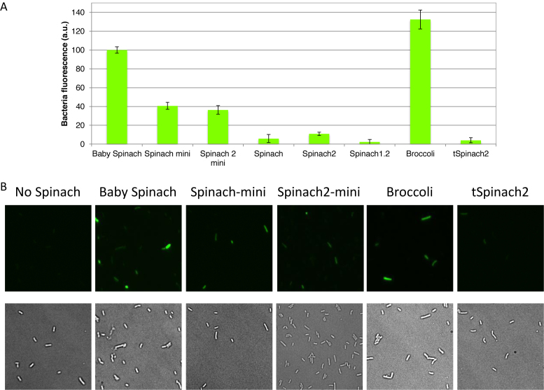
Figure 7.
In vivo fluorescence of cells expressing Spinach variant 16S rRNAs. (A) Fluorescence signal from E. coli TA531 cells incubated with 20 μM DFHBI-1T in M9 (2 mM Mg2+) buffer at 37°C for 45 min. Samples were excited at 470 nm in a plate reader, and fluorescence was recorded at 525 nm. Baby Spinach signal was set as 100. Error bars are s. e. m. for three independent experiments. (B) Imaging of E. coli TA531 cells expressing Spinach-variant-tagged or Broccoli-tagged ribosomes. Cells we incubated with 200 μM DFHBI-1T for 90 min at 37°C, mounted on an agar pad and imaged at room temperature under a microscope. Brightness of the images was adjusted based on Baby Spinach signal. Note heterogeneity in the levels of fluorescence at the single-cell level.

