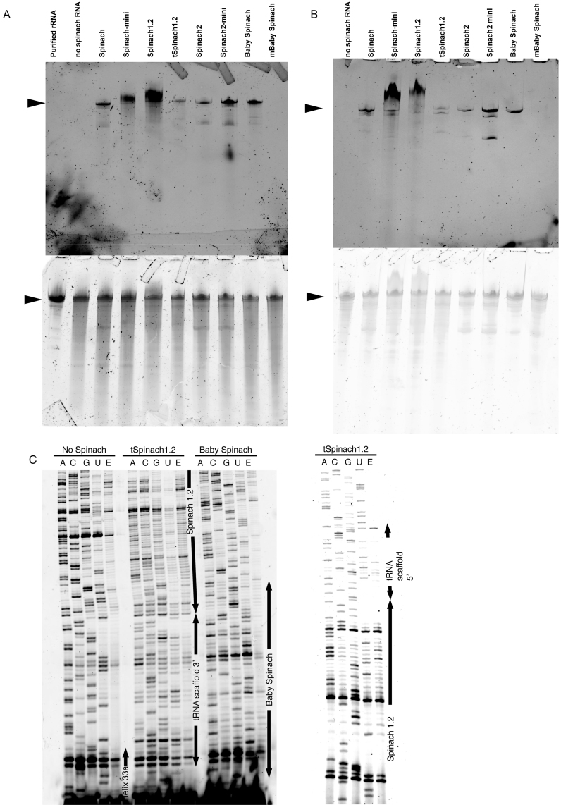
Figure 8.
Degradation of Spinach-tagged 16S rRNA. (A) Total RNA from E. coli TA531 cells expressing Spinach-tagged ribosomes was separated on urea-PAGE and stained with DFHBI-1T (top) and SYBR Gold (bottom). 16S rRNA bands are indicated by arrows. (B) Ribosomes were purified from E. coli TA531 cells expressing Spinach-tagged 16S rRNA. RNA was separated on urea-PAGE and stained with DFHBI-1T (top) and SYBR Gold (bottom). (C) Fluorescent primer extension analysis of cleavage sites in Spinach-modified 16S rRNA. Fluorescent primer extension products generated using Cy5-labelled DNA primer were analysed on urea-PAGE. Regions corresponding to helix 33a, tRNA scaffold, Spinach and Baby Spinach are indicated by arrows.

