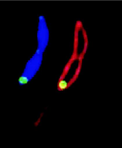
FIG. 5.
Localization of PleD in C. crescentus. Subcellular localization of PleD* (a constitutive active form of PleD) at the stalked pole of a Caulobacter predivisional cell. Green, PleD*-GFP; red, membrane (stained with FM4-64); blue, DNA (stained with DAPI [4′,6′-diamidino-2-phenylindole]). (Figure kindly provided by Urs Jenal.)

