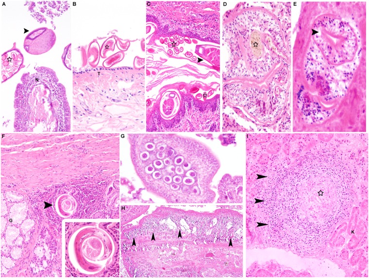
Fig 4. Parasites in tissue sections.
Manifestation of Capillaria infection in various tissues in foxes. (A) Nasal mucosa (N), fox. Mild, diffuse, lymphoplasmahistiocytic inflammation. Within the lumen of the nasal cavity, there are adult nematodes (arrowhead) with a prominent stichosome and a large number of oval, embryonated eggs (star), morphologically consistent with Capillaria sp (HE x100). (B) Trachea, fox. Capillaria eggs (star) are also present on the surface of the tracheal mucosa (T; HE x200). (C) Anal sac, fox. The epithelium is markedly thickened and expanded by a large number of nematode stages, adult nematodes (arrowhead) as well as nematode eggs (asterisk) embedded in the stratum corneum (HE x100). Manifestation of trematode infection (D, E) Lung, raccoon dog. Single adult trematodes with brownish pigment (D; asterisk) and a sucker (E, arrowhead) resembling Alaria alata. There is a lack of pronounced inflammatory response to these parasites (HE x200). (F) Stomach, raccoon dog. Focal, granulomatous inflammation beneath gastric glands (G) with an intralesional nematode parasite (arrow), morphologically consistent with Ollulanus tricuspis (HE x100). Inset: Higher magnification of the nematode (HE x400). (G, H) Intestine, fox. Presence of cestodes in the intestinal lumen (HE x200). (H) Note numerous calcareous corpuscules in the cestode organism resembling Taenia sp. (arrowheads; HE x40). (I) Kidney, raccoon dog. Granulomatous nephritis. Centrally, there is evidence of a necrotic parasite, presumably larval stages of Toxocara (asterisk; HE x100). HE, hematoxylin and eosin.

