. 2017 Apr 11;6:e22706. doi: 10.7554/eLife.22706

Figure 4. Knock down of mpc1 or apc leads to altered mitochondrial respiration and pyruvate metabolism.

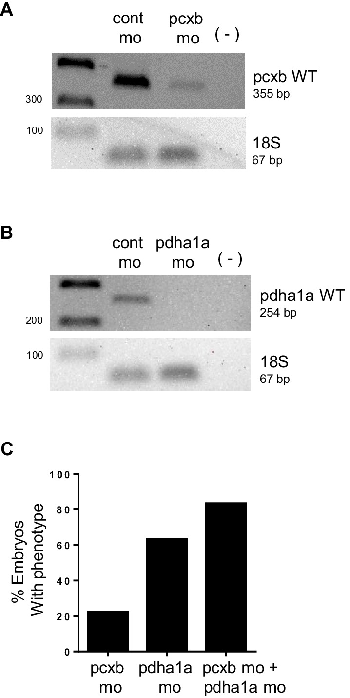
(A) Mitochondrial respiration was evaluated by measuring oxygen consumption rates (OCR) in 72 hpf embryos. (B) Triglyceride (TG) levels were determined in lysates prepared from 72 hpf embryos using a colorimetric assay. (C,D) Quantitative RT-PCR analysis of enzymes involved in pyruvate metabolism in apc mo and (C) apcmcr (D) embryos. pyruvate dehydrogenase alpha 1a (pdha1a); pyruvate dehydrogenase kinase, isozyme 1 (pdk1); pyruvate kinase, muscle, a (pkma); citrate synthase (cs). (E) Lactate levels in apc wild type (WT), un-injected apcmcr (UI) or apcmcr embryos injected with human MPC1 mRNA (MPC1 RNA). For figures A–E, values represent mean ± SD. Graph shown above is representative of at least three independent experiments. Statistical significance was analyzed using unpaired t-test. (F,G,H) Gross phenotype (F), alcian blue staining (G) and in situ hybridization for fabp2 (H) in pdha1a, pcxb, and pcxb + pdha1a mo. pcxb (pyruvate carboxylase b). See also Figure 4—figure supplements 1, 2.

DOI: http://dx.doi.org/10.7554/eLife.22706.010

Figure 4—source data 1. Mean and standard deviation values for Figure 4A,B,E; fold change calculations for Figure 4C,D.
DOI: 10.7554/eLife.22706.011

Figure 4.

Figure 4—figure supplement 1. Knockdown of mpc1 leads to dysregulated pyruvate metabolism.

Figure 4—figure supplement 1.

(A) A simplified scheme showing key enzymes (in blue) involved in pyruvate transport and metabolism (adapted from Schell and Rutter [2013]). (B) Quantitative RT-PCR analysis of pyruvate metabolism enzymes in mpc1 mo. (C) Quantitative RT-PCR analysis of mpc1 in mpc2 mo. For B–C, values represent mean ± SD. Graph shown above is representative of at least three independent experiments. Statistical significance was analyzed using unpaired t-test. pyruvate dehydrogenase alpha 1a (pdha1a); pyruvate carboxylase b (pcxb); lactate dehydrogenase A4 (ldha); pyruvate dehydrogenase kinase, isozyme 1 (pdk1); citrate synthase (cs); solute carrier family 16, member 1 (slc16a1); solute carrier family 16, member 3 (slc16a3); pyruvate kinase, muscle, a (pkma); mitochondrial pyruvate carrier 2 (mpc2).
Figure 4—figure supplement 1—source data 1. Fold change calculations for Figure 4—figure supplement 1B,C.
DOI: 10.7554/eLife.22706.013
Figure 4—figure supplement 2. PCR analysis confirming knockdown of pdha1a, pcxb.

Figure 4—figure supplement 2.

(A,B) cDNA from 48 hpf embryos injected with cont mo, pcxb mo or pdha1a mo was used to amplify a band corresponding to pcxb WT (355 bp) (A) or pdha1a WT (254 bp) (B). A negative control with no reverse transcriptase (-) was also included. Amplification of 18s served as control for input cDNA (67 bp). (C) Microinjection of WT embryos with pdha1a morpholino (pdha1a mo) resulted in a higher penetrance compared to pcxb morpholino (pcxb mo). Co-injection of both morpholinos gave the highest percentage of embryos with phenotype (pcxb mo + pdha1a mo).