. 2017 Mar 7;6:e23661. doi: 10.7554/eLife.23661

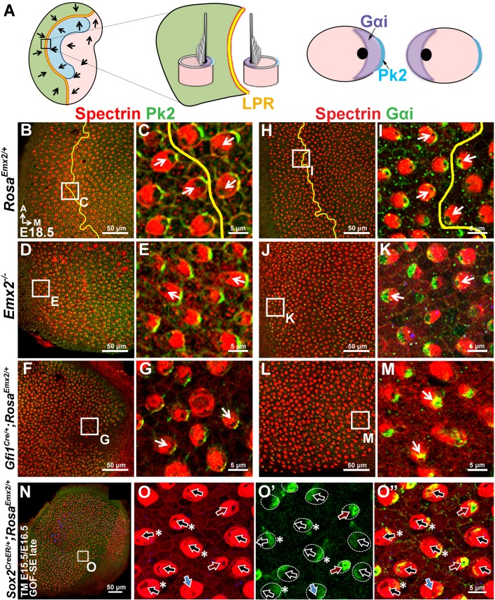
Figure 6. Hair bundle polarity reversal is associated with reversed Gαi but not Pk2 localization in Emx2 mutants.

(A) Schematic diagram illustrates the distribution of Pk2 and Gαi in HCs across the LPR of a control utricle. (B–C) In control utricles, Pk2 staining (green) is concentrated on the medial side of HC-supporting cell border across the LPR (n = 15). (D–G) In the lateral region of Emx2-/- (D-E; n = 5) and medial region of Gfi1Cre/+;RosaEmx2/+ utricles (F-G; n = 4), in which the hair bundle polarity is reversed compared to the respective left and right side of the LPR in controls (C), Pk2 staining remained on the medial side (green). (H–M) In control utricles (H–I), Gαi immunostaining (green) is associated with the kinocilium. This relationship is consistent among all HCs of the utricle and the staining pattern of Gαi is reversed across the LPR (yellow line) (I; n = 8). A similar spatial relationship between Gαi and the kinocilium position is observed in Emx2 -/- (J-K; n = 4) and Gfi1Cre/+;RosaEmx2/+ (L-M; n = 5) utricles as controls. (N–O”) Sox2CreER/+;RosaEmx2/+ utricles ( GOF-SE late) show mixed phenotypes including normal (black arrows), misoriented (blue arrows), and reversed (red arrows) kinocilia (n = 3). Some HCs with normal polarity show the abnormal Gαi distribution (asterisks).

DOI: http://dx.doi.org/10.7554/eLife.23661.016

Figure 6.

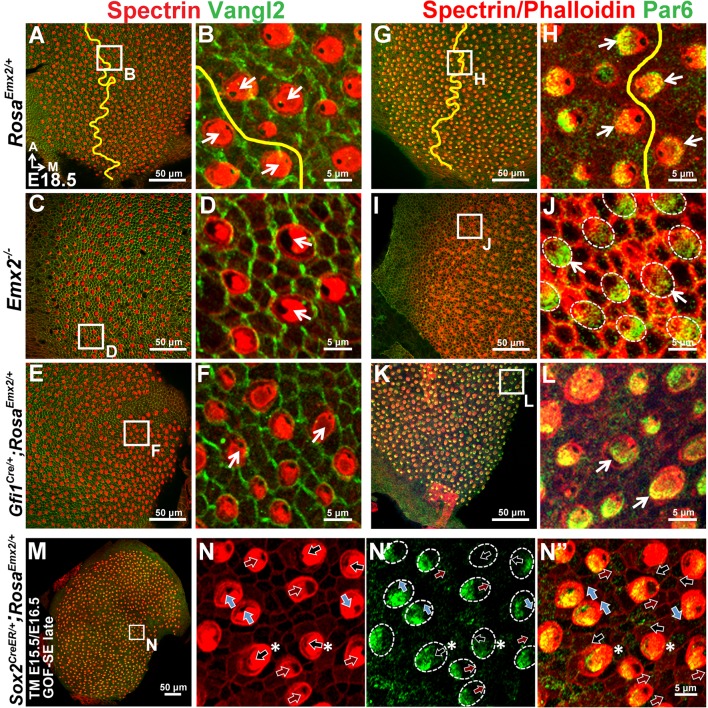
Figure 6—figure supplement 1. Distribution of Vangl2 and Par6 immunoreactivites in apical HCs is not changed in Emx2 mutant utricles.

Figure 6—figure supplement 1.

(A–B) In controls (n = 3), Vangl2 immunostaining (green) are restricted, stronger between adjacent supporting cells and weaker between HC and supporting cell junction. The staining pattern of Vangl2 is not changed across the LPR (Jones et al., 2014). (C–F) In the lateral region of Emx2-/- utricle (C-D; n = 3) and medial region of Gfi1Cre/+;RosaEmx2/+ utricle (E-F; n = 3), in which the hair bundle polarity is reversed compared to controls (B), there is no obvious change in the distribution of Vangl2 immunostaining. (G–L) In control utricles (G-H; n = 3), Par6 (green) is localized in the apical surface of HCs opposite to the kinocilium (H). This relationship between kinocilium and Par6 is consistent in all HCs and the staining pattern of Par6 is reversed across the LPR (yellow line). Similar relationship between Par6 immunostaining and kinocilium position (lack of phalloidin staining) is observed in Emx2 -/- (I-J; n = 3) and Gfi1Cre/+;RosaEmx2/+ (K-L; n = 3) utricles as controls. (M–N”) Activating Emx2 at a later age by administering tamoxifen at E15.5 and E16.5 causes mixed phenotypes in Sox2CreER/+;RosaEmx2/+ utricles (GOF-SE late; n = 3): normal (black arrows), reversed (red arrows) or misoriented (blue arrows) kinocilia. In some HCs with normal hair bundle polarity, anti-Par6 distribution is no longer complementary to the kinocilium position (asterisks).
Figure 6—figure supplement 2. Distribution of Pk2 immunoreactivites in apical HCs is maintained in Emx2 GOF-SE late mutant utricles.

Figure 6—figure supplement 2.

In the GOF-SE late utricle (A–B"), hair bundles can be normal (black arrows), reversed (red arrows), or misoriented (blue arrows). However, the immunoreactivities of Pk2 is maintained on the medial side of HC-supporting cell boundary regardless of the hair bundle polarity.