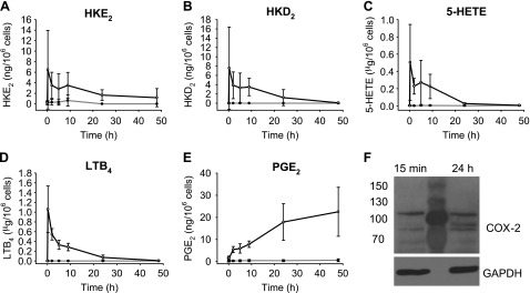
Figure 3.
Time course of eicosanoid biosynthesis in human leukocytes. A–E) HKE2 (A) and HKD2 (B), as well as 5-HETE (C), LTB4 (D), and PGE2 (E) were quantified using LC-SRM-MS analyses after derivatization with AMPP. F) Western blot analysis of COX-2 expression in leukocytes at 15 min and after 24 h stimulation with LPS. The central lane was loaded with protein molecular mass markers. Leukocytes were isolated from peripheral blood and stimulated with LPS (10 μg/ml) at 37°C for the time indicated (15 min, 2, 5, 9, 24, and 48 h) including treatment with A23187 (5 μM) during the final 15 min. Open circles: LPS-treated samples; closed circles: vehicle treated (unstimulated) controls. Error bars: SD of leukocyte samples obtained from 3 volunteers.

