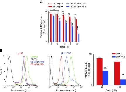
Figure 6.
Effects of pHK and pHK-PAS on intracellular ATP levels and mitochondrial membrane potential. A) Dose- and time-dependent decrease in intracellular ATP levels. HeLa cells were treated with pHK (red bars) or pHK-PAS (blue bars) for the indicated durations. Two peptide concentrations, 25 (open bars) or 50 µM (filled bars), were used. ATP levels were then measured by using the CTG assay. Cells that were treated with peptide-free carrier were used as control. Relative ATP levels were determined from the ratio of the luminescence of treated cells to control cells. ns, nonsignificant (P > 0.05). **P < 0.001, ***P < 0.0001 compared with controls. B) ΔΨm depolarization of the inner mitochondrial membrane. HeLa cells were treated with 25 or 50 µM pHK (left) or pHK-PAS (middle) for 24 h, and fluorescence of the ΔΨm probe TMRM was measured by using FACS. Cells that were treated with the uncoupling agent, FCCP (10 µM), were used as positive controls for ΔΨm dissipation, and cells that were treated with vehicle alone served as negative controls. TMRM fluorescence intensity is plotted as a percentage of controls (right). **P < 0.001 compared with pHK.

