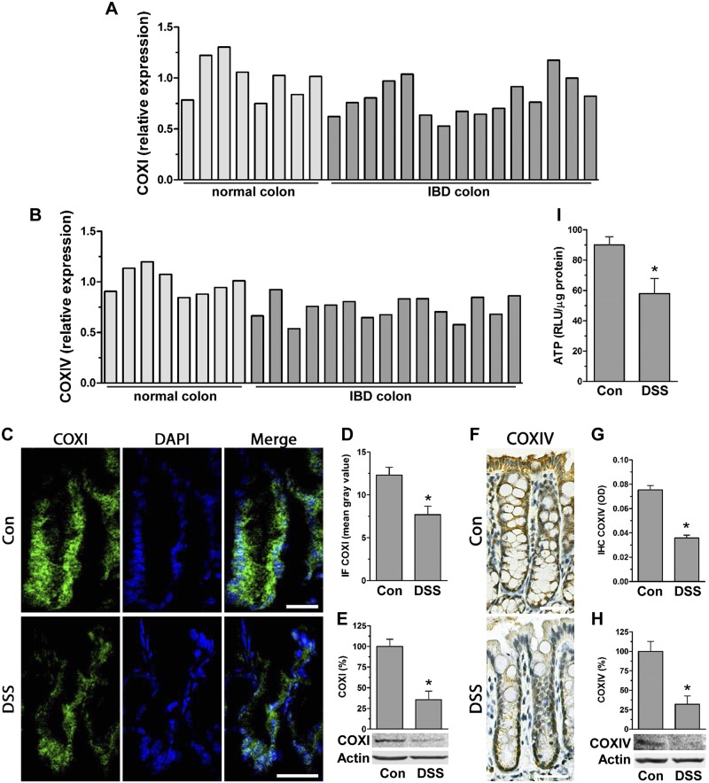
Figure 1.
Intestinal inflammation is associated with decreased levels of mitochondrial complexes COXI and IV. A, B) Microarray relative expression of COXI (A) and COXIV (B) in human IBD colon (GSE4183) (normal colon, n = 8; IBD colon, n = 15). P < 0.05, IBD compared to normal colon, Student’s t test. C, D) Frozen colon tissue from DSS-treated mice was immunofluorescently stained for COXI (Alexa Fluor 488; n = 4 mice). Scale bars, 40 μm. Graph shows quantification of fluorescence displayed as mean gray value (ImageJ; n = 10 crypts.) *P < 0.05 compared to control (con), Student’s t test. E) Total protein of scraped mucosa from con and DSS-treated mice was immunoblotted for COXI (densitometric analysis of n = 3). *P < 0.05, compared to con, Student’s t test. F, G) Paraffin-embedded colon tissue of mice treated with DSS was immunohistostained for COXIV (n = 4 mice). Scale bars, 40 μm. Graph represents quantification of DAB staining expressed as optical density (OD) (ImageJ; n = 20 crypts). *P < 0.05 compared to con, Student’s t test. H) Total protein extracted from scraped mucosa of DSS-treated mice was immunoblotted for COXIV (densitometric analysis of n = 3). *P < 0.05, compared to con, Students t test. I) ATP levels [relative luciferase unit (RLU)/µg protein] were quantified by luciferase driven bioluminescence in scraped mucosa of DSS-treated mice (n = 6). *P < 0.05, compared to con, Student’s t test.

