Figure 6.
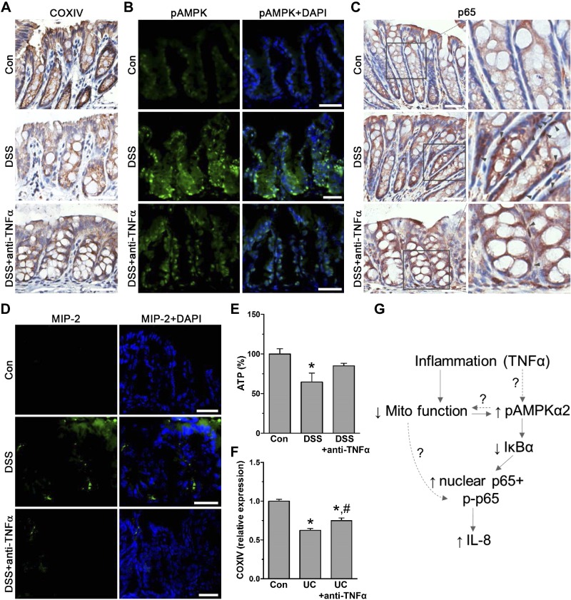
In human and mouse colon, anti-TNF-α treatment restores reduced mitochondrial dependent inflammation. A) Immunohistostaining for COXIV in paraffin-embedded colon tissue from control (con) and DSS-treated mice with or without anti-TNF-α (n = 4 mice). Scale bars, 40 μm. B) Immunofluorescence staining of frozen colon tissue for pAMPK of con and DSS-treated mice with or without anti-TNF-α (Alexa Fluor 488; n = 4 mice). Scale bars, 20 μm. C) Immunohistostaining for NF-κB subunit p65 of paraffin-embedded colon tissue from con and DSS-treated mice with or without anti-TNF-α. Magnified area shows nuclear p65 localization (gray arrows; n = 4 mice). Scale bars, 40 μm. D) Immunofluorescently stained frozen colon tissue of DSS and anti-TNF-α-treated mice for IL-8 ortholog MIP-2 (Alexa Fluor 488; n = 4 mice). Scale bars, 20 μm. E) ATP levels in scraped mucosa of con, DSS, and anti-TNF-α-treated mice (n = 6). *P < 0.05, compared to con, ANOVA. F) Microarray expression of COXIV relative to con (n = 6) in human ulcerative colitis (UC) biopsy samples before (n = 24) and 6 wk after anti-TNF-α treatment (n = 24; GSE16879). *P < 0.05, compared to con; #P < 0.05 compared to UC alone, ANOVA. G) Proposed regulatory mechanism of colon inflammation dependent on reduced mitochondrial function leading to AMPKα2 activity.

