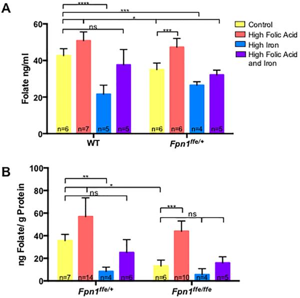
Figure 4. Folate levels in dams and embryos.
A. Determination of red blood cell folate levels in pregnant dams. Whole blood was obtained from pregnant wildtype (WT) or Fpn1ffe/+ dams at 9.5 or 11.5 dpc. Dams were supplemented with control (yellow bar), high folic acid (10 ppm, orange bar), high iron (0.5% carbonyl iron, blue bar) or high folic acid and iron (purple bar) diets for 4 weeks before mating. B. Determination of folate levels in 11.5 dpc wildtype (Fpn1+/+) and Fpn1ffe/ffe embryos from dams fed the various diets. The number of samples represented in each group is indicated. The Sidak's test was used to determine significance of multiple comparisons within a genotype and the Tukey test across genotypes. P-values: ≤0.05 *, ≤0.01**, ≤0.001 ***, ≤0.0001 **** or non significant (ns). The number of samples represented in each group is indicated.

