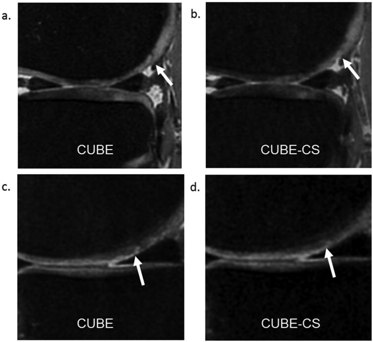
Figure 6.

(a and b) CUBE and CUBE-CS images in a 37 year old male show decreased conspicuity of a dark linear cartilage fissure on the posterior medial femoral condyle (arrows) on the CUBE-CS image due to increased image blurring. (c and d) CUBE and CUBE-CS images in a 41 year old female show decreased conspicuity of a superficial partial-thickness cartilage lesion on the central medial femoral condyle (arrows) on the CUBE-CS image due to increased image blurring.
