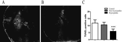
Fig. 2.
Effects of DBP on apoptosis in the GTs from male fetuses at GD 19. Apoptotic cells (red dots, 20×) in the GT in the control (A) and hypospadiac (B) rats detected by TUNEL. (C) Quantitative analysis of the number of apoptotic cells in the GT. Three high power fields per section were randomly selected and at least 1,000 cells per field were counted on coded sections in a blind manner (n=18, from 6 sections). ***, p<0.001.

