
Fig. 1.
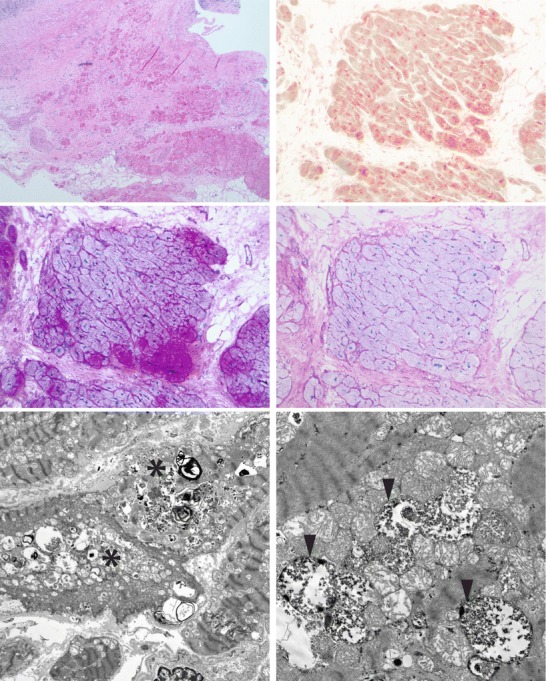
Histologic and ultrastructural features of myocardial tissue taken during LVAD placement, showing features of a lysosomal glycogen deposition disorder. Top row, left: paraffin-embedded H&E section showing extensive fibrosis and myocyte loss, myocyte hypertrophy, and vacuolization, with reactive, pericardial, mononuclear inflammation. Top row, right: frozen section stained for acid phosphatase activity showing diffusely increased lysosomal activity. Middle row, left: PAS staining of frozen section showing increased glycogen in myocytes, in an uneven pattern. Middle row, right: PASD (frozen) showing only minimal diastase-resistant material. Bottom row, left: electron microscopic image at 2200× showing vacuoles (asterisks) containing glycogen and lysosomal structures. Bottom row, right: electron microscopic image at 9300× showing that the glycogen is frequently membrane-bound (arrowheads)