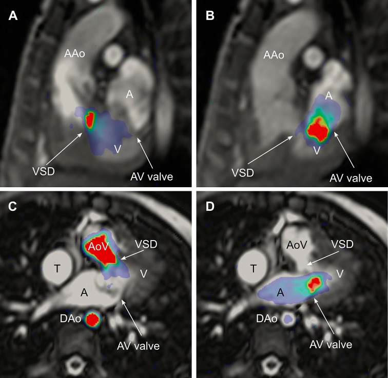
Fig. 2.
Ventricular kinetic energy (KE) in a Fontan patient superimposed on CMR images to visualize the anatomical location of KE. The upper panel shows an oblique sagittal view in systole (a) and diastole (b). Systolic peak KE can be seen in a ventricular septal defect leading the blood to the aorta. Diastolic peak KE is located from the atrioventricular valve into the ventricle. The lower panel visualizes KE in an oblique transversal view in the same patient in systole (c) and diastole (d). AAo ascending aorta; VSD ventricular septal defect; V ventricle; A atrium; AV valve atrioventricular valve; AoV aortic valve; DAo descending aorta; T lateral tunnel

